Our Services

-
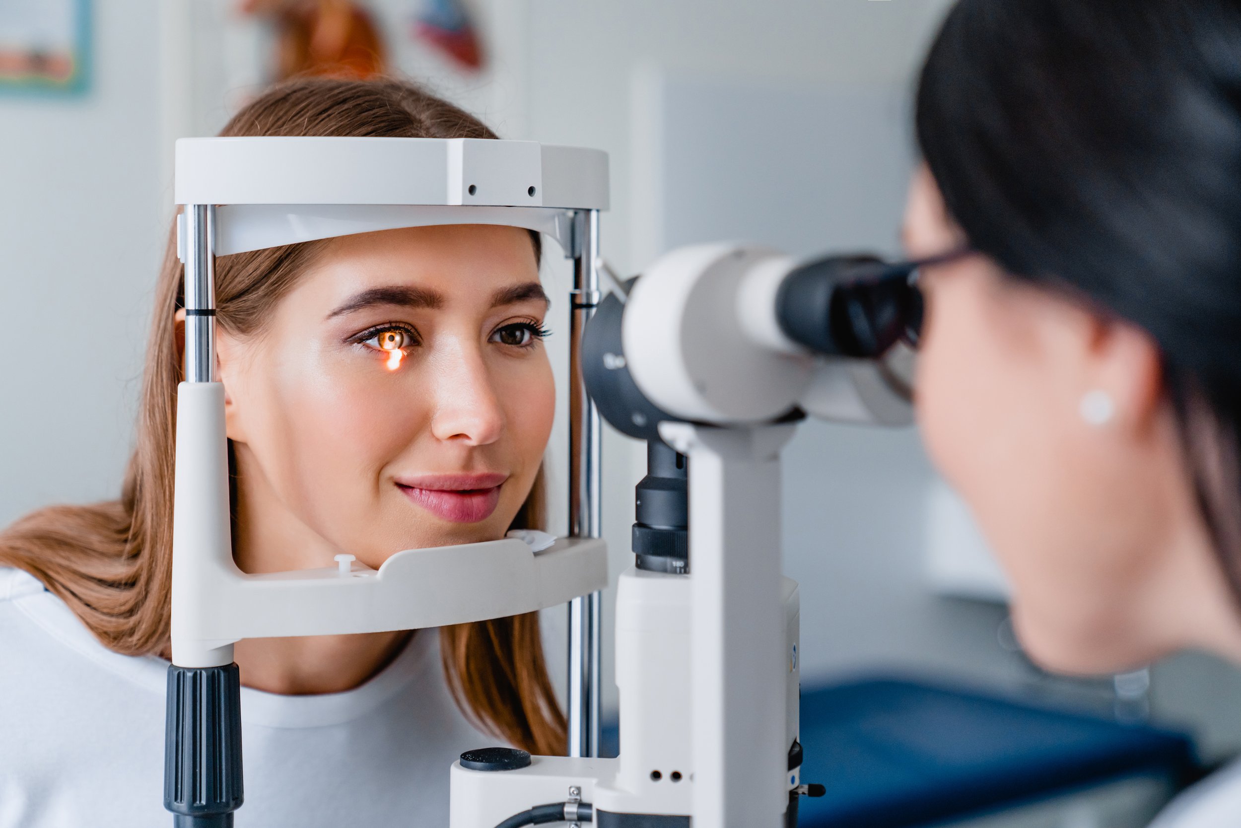
An eye examination consists of a series of tests which are performed to both assess your vision and to enable the detection of eye disease and ocular manifestations of systemic disease. The practice is equipped with the latest diagnostic instrumentation and the examination will be tailored to your individual needs. Following your examination you will be advised whether your sight needs correcting and about any other issues relating to the health of your eyes.
The aim of visual correction is to optimise both clarity of vision and overall visual comfort. This involves not only correcting focusing errors but also any other anomalies such as muscular imbalances and poor eye co-ordination. Should a spectacle correction be required your optometrist will lead you through the dispensing component so as to enable you to choose the most suitable spectacle frame and lenses for your individual requirements.
-
Optical Coherence Tomography is a highly sophisticated procedure whereby three-dimensional, cross sectional scans of the eye are taken. It provides high resolution images of the anterior part of the eye, the vitreous, retina, macula, optic nerve head and choroid. It is the only technique available which resolves the substructure of the retina in living eyes.
The scan is non-invasive, painless, simple and quick and is a very advanced screening system that checks for potentially serious eye diseases. It is useful in the diagnoses of diseases, monitoring the progression of disease and assessing the response to treatment. Some common conditions identified with regular OCT screening include age-related macular degeneration (AMD), glaucoma, diabetic retinopathy and diabetic maculopathy, vitreous detachments, epiretinal membranes and macular holes.
OCT in glaucoma
Glaucoma is one of the leading causes of blindness. Worldwide it affects up to 2% of people over the age of 40 years, and up to 10% over the age of 80.
It is a progressive disease of the eye which results in damage to the optic nerve and thinning of specific layers within the retina. Damage occurs over a period of time and as it progresses patients may experience an increasing loss of their vision. Unfortunately any loss of vision which occurs is irreversible.
There is no guaranteed protection against glaucoma however early detection may help protect from the severe consequences of this disease.
OCT scanning takes a series of three-dimensional scans of the optic nerve head and the central retina. A computer compiles and analyses the information from the scans and produces a "baseline" image. Comparing future scans over time enables even the smallest and earliest changes to be detected.
-
OCT-A is an imaging modality which provides detailed visualisation of the perfusion of vascular networks in the eye. It enables us to visualise the retinal microvasculature and blood flow in different individual segments of the retina and choroid. ie. how much nutrition is getting to the retina and choroid.
It enables detection of early changes in the retinal and choroidal blood flow in a number of eye conditions such as age-related macular degeneration, glaucoma, diabetic retinopathy, retinal vein occlusions, central serous chorioretinopathy, retinal pigment epithelial detachments and optic neuropathies.
OCT-A in diabetic retinopathy
OCT-A can detect vascular changes and abnormalities in diabetic patients even before the normal retinal signs of diabetic retinopathy are present.
-
Digital retinal imaging enables us to take photographs and record in detail the appearance of the back of the eye. This provides us with an accurate method of screening for eye disease and by comparing photographs over time enables us to monitor for and detect any subtle changes at the back of the eyes.
-
A visual field examination is a test used to determine the sensitivity of the eye and whether there are any abnormalities in a person's central or peripheral vision. These may be caused by various eye diseases as well as underlying medical conditions such as brain tumours or a stroke.
Examinations are conducted using computerised screeners including the Humphrey visual field analyser. This has become an industry standard perimeter for the diagnosis and monitoring of glaucoma and other ocular and neurological diseases.
-
We offer a diverse selection of spectacle frames from designer brands and other high quality ranges to more affordable items. Frames of innovative style and high quality craftsmanship.
The look, quality and wearing comfort of spectacles is important and our experienced staff will help and advise on the suitability and functionality of a frame combined with prescription compatibility. We dispense and supply spectacle lenses from the world’s leading manufacturers such as Zeiss, Hoya and Essilor.
-
More people are discovering the benefits and freedom of contact lenses. Whether it be for full time wear or just for sports or social wear, there are now lenses to suit almost anybody. We fit a wide range of contact lenses from the world's leading manufacturers. Our aim is to find the most suitable type of contact lens for each patient.
All new patients can expect to undertake a comprehensive trial period during which their compatibility to the lenses they are trialling is determined. This will usually take a few weeks and may involve trying a few different types of contact lenses to find which lenses are best for them.
We have considerable experience at fitting even the most complex cases and from the initial consultation through to regular aftercare checks, all appointments are carried out by our optometrists.
-
Eye examinations for children are extremely important. A child's vision develops until around the age of 7-8 and it is essential during these early years that all is progressing normally.
Children will often not complain of any difficulty, especially if only one eye is affected. Early identification of a problem is crucial as children are more responsive to treatment when problems are identified early. Should a child require spectacles or have a squint which is left untreated, they may have permanently reduced vision (amblyopia).
It is recommended that children have their first examination at 12-18 months unless there is a suspected problem. We will then continue to monitor their progress at regular intervals.
Parental involvement is encouraged during the examination and all findings will be discussed with you.